IMPORTIR & DISTRIBUTOR ALAT LABORATORIUM & ALAT KESEHATAN
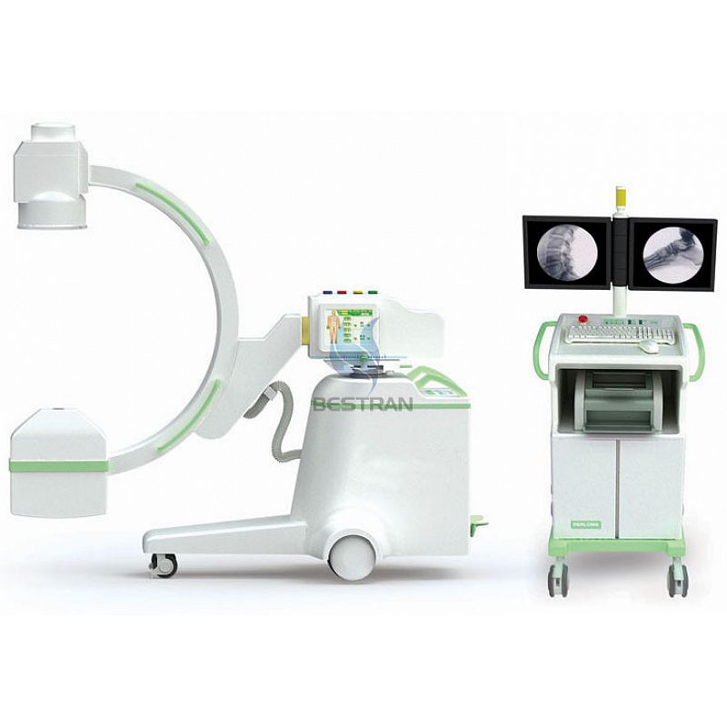
BT-XC07 is wildly used in Department of Orthopedics, Department of General Surgery, orthopedic surgery department, Urology Surgery, Spine Surgery, Abdominal Surgery, Department of Pain Treatment, Cardiology, Gastroenterology, Gynecology, Operating Room, etc.
2 Intelligent control of exposure, achieve low radiation dose.
3 Multiple operating modes to meet various clinical needs.
4 Muti-leaf and vertical light shadow control, effectively, reduces soft X-ray ,dramatically reducing the skin dose.
5 Imported famous brand image intensifier and all-digital CCD camera providing high-quality and high-resolution images.
6 Dual high-resolution LCD monitors assure image quality effect.
7 The powerful digital graphic workstation standard configuration DICOM 3.0 interfaces and network perfect butt, support Worklist registration and manual registration dual registration modes
.8 Workstation has the high-capacity digital storage function. Fluoroscopy and digital spot film(DSI) are all stored in digital format without any loss. It has powerful processing capabilities like edge enhancement, multiple images, gamma correction,movie playback, window width/level, experts template mode, burn CD, etc.
9 Four-D electric motion control, accurate positioning, flexible and comfortable control. Super large frame design, provides lager diagnostic and watch spaces and more comfortable surgical operation environment. New design and new ideas brings superior experience.
10 Two panels of human graphical LCD touch screen provide intelligent ,fast and convenient operation. Dual motion control system and double foot brake for expose make this machine greatly meet the demands of clinical operations.
3 Configurations
(6.0kW, 60kHZ, 125kV) 1set
8.19 inch LCD display 2sets
| Category | Items | Content |
| Electrical Performance | High frequency inverter power supply | Power output: 6.0kW; Main Inverter Frequency: 60 kHz |
| Automatic &Manual Continuous Fluoroscopy | Tube voltage:40kV~125kV continuously adjustable | |
| Tube current:0.3mA~4mA continuously adjustable | ||
| Automatic &Manual Enhanced Fluoroscopy | Tube voltage:40kV~125kV continuously adjustable | |
| Tube current:0.3mA~8mA continuously adjustable | ||
| DSI Digital Spotfilm Imaging | optional 1-5 fps | |
| Photography tube KV ,mA | 40kV~125kV 120mA | |
| X ray tube | X-ray tube special for high frequency | Rotary anode focus 0.3/0.6 mm |
| Anode thermal capacity:212kJ | ||
| Imaging
system |
Image Intensifier | Toshiba 9″ image intensifier |
| CCD camera | Medical million-pixels ultra-low illumination digital camera | |
| CCU | Real-time acquisition、up and down image, left and right mirror image、continuous adjustable recursive noise reduction、multy images storage、image patching,、LIH (last image freeze) | |
| Monitor | 19 inch LCD display | |
| Workstation software | Storage without loss,multi-image display, Image W/L real-time adjustment, grayscale conversion, interest area balance, Gamma correction, reversal, noise reduction,enhancement,smoothing,sharpening,compression,enlargement,measure, mark, image & document report printing& typeset,Expert template, Dicom image sending, Dicom image print, movie playback, image burn record, worklist registration, etc | |
| Structure | Direction wheel and main wheel | Direction wheel can rotate in any direction,main wheel±90° |
| C-arm | Forward & backward 200mm by electrical power Revolution around horizontal axis : ±180°. Revolution around vertical axis: ±15° | |
| Slipping on orbit: 120°(+90°~-30°) electromotion ,The pillar up and down electromotion is 400mm. | ||
| SID:1060 mm C-arm open :860 mm Arch depth:700 mm |
Tidak ada produk berkaitan.
Tidak ada produk popular.
Format Pemesanan Via SMS
Kirim format SMS di bawah ke 08987654321
#Nama #Alamat Lengkap #Kode Produk #Jumlah Produk #Ukuran Produk
Selanjutnya kami akan membalas SMS anda dengan rincian total belanja anda yang harus ditransfer ke rekening kami
Harap segera konfirmasi jika pembayaran sudah ditransfer ke rekening di bawah ini.
