Rp
Features
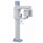
- BT-XD01 Panoramic Digital system is a kind of oral X-ray digital tomography System, it dedicated to the oral and maxillofacial area imaging, and it takes use of the “Tapered beam” Scanning three-dimensional imaging technique.
- BT-XD01 is used for getting the high contrast geometric information in the maxillofacial area during diagnostic process, and it can be used for:
- Mandible / frontal imaging
- Nose surface/ fistula and maxillofacial imaging
After Three-dimensional reconstruction, it can produce slice group and build the Three-dimensional reconstruction model. After this progress, axial slice group constituted volume data. With these data, the machine can real-time display the coronal and sagittal view of the reconstruction model. After defining the area of interest, users can create research data with the volume data. The area of interest can keep the tilt angle with the volume data to get vertical image and to correct the positioning error.
- The equipment could create panoramic, axial surface, and 3D images according to the data research, and it has the function of distance measurement, angle measurement, mark and so on.
- The equipment could construct the scanning objects at random, no need to reconstruct along axis, and no need to reformat the data.
- The equipment has advantage of construction and maintenance, while using the same scanning time, it requests lower output power of x-rays & x-ray tube & scanning device.
- Images could be printed and stored as test reports.
- I. Application
BT-XD01 is widely used for dental CT diagnostic with 12cm*15cm flat panel detector, mainly for oral and maxillofacial surgeries, orthognathic surgeries, plant department and other dental department in hospitals.
- II. Technical Specifications
- Generator
- Anode Voltage: 60-92 kV
- Anode current: 1-15 mA
- Max.output power: 1.38kW
- Parameter range: 60-92KV, 1-15mA
- Information display: kV, mA, Ascending &Descending position, Laser indication, human features
- X-ray Tube
- Model: D-054SB (Toshiba)
- Focus: 0.5mm
- Thermal capacity: 35kJ (50kHU)
- Radiography mode: Cone Beam CT
- Imaging System THALES Flat Panel Detector
- Size: 12cm*15cm
- Pixel size: 150um
- 3D reconstruction size: φ80 *90 mm
- Resolution: 1280 *1024
- Scanning angle: 200°
- Scanning time: 18s
- Exposure time: 4-8s
- Scanning frame: 400 Frames
- 3D Reconstruction time: ≤18s
- Volumepixel size: 0.2 mm
- A/D Gray scale: 14bit
- Acquisition mode: Pulse fluoroscopy
- Detector type: CMOS
- Pixel: 960*786
- Image resolution: ≥3.1lp/mm
- Mechanical Performance
- Vertical up & down stroke: 1000mm
- Noise for ascending & descending:<70dB
- Anticollision: Yes
- Weight: 260kg
- Appearance size: 788*1090*2200mm
- Power supply: 220V, 50/60Hz, 2kVA
- Radiography room requirements: ≥5m2, 2mm lead equivalent