
IMPORTIR & DISTRIBUTOR ALAT LABORATORIUM & ALAT KESEHATAN
Feature
1.Amorphous silicon flat detector, use the advanced manufacturing technology with super stability
2.The detector can move with large scale of 17”×17” effective detection area.can meet all kinds of parts to shoot.
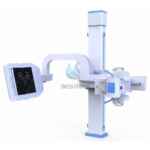
3.The electric lift and rotatable newly designed U-shaped frame can meet the photographic requirements of different standing and lying positions. The U-shaped design can make the operation much more convenient and flexible.
4.The leading domestic high power compact high frequency X-ray generator and high frequency power inverter makes the machine much more compact and more convenient without the extra high-voltage generator and cable.
5.Newly designed photographic bed used specially for the U-shaped arm X-ray machine. The bed floating and electromagnetic lock design makes it convenient for the accurate position of the lying patient.
6.Specially designed working station for DR adopts all-digital intelligent touchable LCD control system which is graphic and real color. This system makes the operation easier and more convenient.
7.Apply different photographic parameters according to the human characteristics, such as multi-site, multi-position, muti-body shape, adult and children etc. The parameters can be modified and stored at will and make the operation more convenient.
8.The high quality high frequency high-voltage X-ray generator and high frequency power inverter can produce high-definition and good contrast images by high quality radiation and low dose.
9.The application of the KV and MA digital closed-loop control technology and the real time control of the microprocessor ensure the accuracy and repeatability of the dose.
10.It provides more than 1200 APR preset program
11.Support Dicom 3.0 convenient to connect with PACS system to print and transmission.
12.Two control modes for Mechanical movement: close-table control, hand-remote control, Cell compartment control, it can easily, flexibility and quickly operated.
13.Image Workstations
Image processing system: Photo processing software, X-ray synchro Control software, motion control software Image post-processing: tissue equilibrium, W/L adjustment,Gamma correction, interest district, reversed phase, noise reduction, smooth, sharpen,pseudo color, Edge extraction, shadow compensation, filter neuclear, single window,dual-window, four windows, movement, right rotated 90°, left rotated 90°, level mirror image, vertical mirror image, magnifying glass, image zooming, reset, layer information, label character, drawing label, length measurement, angle measurement, rectangular length, rectangular area, elliptic length, elliptic area
I. Application:
This machine is applied to take radiography on every part of human body, such as head, limbs, chest, limbus and abdomen and etc.
| High-
frequency X-ray machine |
Output power | 85kW | |
| Main inverter frequency | 260kHz | ||
| X-ray tube
(IAE) |
Dual-focus X-ray tube | Small focus:0.6 Large focus:1.2 | |
| Output power | 35/85 kW | ||
| Anode Thermal Capacity | 210kJ(300kU) | ||
| Thermal capacity | 1280kJ | ||
| Anode Angle | 12° | ||
| Speed of rotating anode | 10,000rpm | ||
| Tube Current | 10mA- 1000mA | ||
| Tube voltage | 40-150kV | ||
| mAs | 1-1250mAs | ||
| Exposure Time | 0.0005-10s | ||
| AEC | Yes (Option) | ||
|
Digital Image System |
Digital Detector (Thales) |
Field of view | 17”*17” |
| Pixel | 3K*3K | ||
| Ultimate spatial resolution | 3.7LP/mm | ||
| Pixel size | 148um | ||
| Output grayscale | 16bit | ||
| Imaging time | 9s | ||
| Image Workstation | Acquisition module | -Gigabit network acquisition
-Adaptive image enhancement processing |
|
| Image information management | Dicom image transmission
Dicom film printing Dicom image storage (hard disk, compact disk)
|
||
| Mechanical structure and performance | U-arm | Vertical movement range | ≥1250 mm(motorized control) |
| Focus-screen movement range | 1000mm-1800mm(motorized control) | ||
| Rotation range | -40°-+130°(motorized control) | ||
| Detector rotation | 90° | ||
| X-ray tube ratation | 180° | ||
| Photography table
(Optional) |
Table size | 2000mm*650mm | |
| Table height | ≤740mm | ||
| Transverse movement | 200mm(electromagnetic lock) | ||
| Longitudinal movement | 100mm(electromagnetic lock) | ||
| Power supply | 380V 50/60Hz | ||
III. Standard Configurationt
| No. | Item | Quantity |
| 1 | Newly designed U-shaped frame | 1 |
| 2 | X-ray tube assembly | 1 |
| 3 | Combined high-frequency high-voltage X-ray | 1 |
| 4 | 17〞×17〞Digital flat | 1 |
| 5 | 24〞Gray scale medical LCD display | 1 |
| 6 | Colored LCD touch screen control box | 1 |
| 7 | Electric box | 1 |
| 8 | Image post-processor (with image processing software) | 1 |
| 9 | Collimator | 1 |
Tidak ada produk berkaitan.
Tidak ada produk popular.
Format Pemesanan Via SMS
Kirim format SMS di bawah ke 08987654321
#Nama #Alamat Lengkap #Kode Produk #Jumlah Produk #Ukuran Produk
Selanjutnya kami akan membalas SMS anda dengan rincian total belanja anda yang harus ditransfer ke rekening kami
Harap segera konfirmasi jika pembayaran sudah ditransfer ke rekening di bawah ini.
