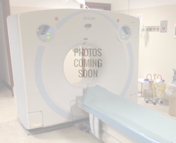
CT SCAN HITACHI SUPRIA 16 SLICE

CT SCAN HITACHI SUPRIA 16 SLICE

5 (1 votes)

share

The need for faster and more accurate diagnosis is increasing every day in front-line medical practice. Supria is designed to…
sold out
Rp


The need for faster and more accurate diagnosis is increasing every day in front-line medical practice. Supria is designed to answer, in one CT, all the demands of diverse routine applications, with the added benefits of a compact size, excellent image quality and ease of use without compromise. Supria CT is your answer to take off to the next clinical and technology standard.
High speed scanning with less than 1 sec/rot, the latest technologies and a 75cm bore, one of the widest among the presently available 16/64 slice CT systems, enable a precise and comfortable examination. Supria achieves the minimum dose and reduces image noise and artifact to provide higher quality images.
Whole body high speed scanning with less than 1 sec/rot
75 cm wide gantry bore with compact foot-print
Effective field of view: 500mm
X-ray tube: 5 MHU
Maximum output: 48kW
Supria is equipped with the latest technologies for low dose imaging and excellent image quality
State of the art technologies for minimum dose are integrated as standard (ALARA = As Low As Reasonably Achievable)
Hitachi’s latest noise reduction technology – Iterative Reconstruction Processing (Intelli IP Advanced) is applied and integrated to achieve low dose and high image quality
Unique 3D reconstruction algorithm (CORE method) ensures high image quality with fewer artifacts even during high pitch scanning
New generator design enables improved power supply efficiency
Open Access and a 75cm wide bore for increased patient comfort
Easy positioning thanks to the open gantry
Intuitive GUI design with Quick-Entry mode allows simple operation for all users
The 24-inch wide monitor clearly displays all necessary information at a glance
Very compact 16/64 slice CT systems thanks to improved gantry design and the need for only 3 system modules; gantry, patient table, and operator console.
Hyper Q-net: image analysis software
FatPointer: Body analysis software
RiskPointer: LAA analysis software
CT Colonoscopy: Colon analysis software
Dental Analysis: Tooth-jaw analysis software
DICOM MWM: Modality Worklist Management
DICOM MPPS: Modality Performed Procedure Step
DICOM Q/R: DICOM Query/Retrieve
Complete 3D reconstruction package
Specifications
Features
Packages
Clinical Images
Coloured 3D Volume Rendering reconstruction of post-contrast brain. Detailed visualization of post-operative left cerebral artery aneurysm.*

Coloured 3D Volume Rendering reconstruction of post-contrast brain. Detailed visualization of post-operative left cerebral artery aneurysm.*
Coloured 3D Volume Rendering reconstruction with cut-plane option allows visualization of the lung fields for pneumonia.*

Coloured 3D Volume Rendering reconstruction with cut-plane option allows visualization of the lung fields for pneumonia.*
Maximum Intensity Projection reconstruction in grey scale for lung pneumonia.*

Maximum Intensity Projection reconstruction in grey scale for lung pneumonia.*
Coloured 3D Volume Rendering reconstruction with fusion images showing arterial and portal phase post contrast abdominal angiography using Intelli EC and PredictScan (Bolus-Tracking). The HCC (hepatocellular carcinoma) is visible in red.*

Coloured 3D Volume Rendering reconstruction with fusion images showing arterial and portal phase post contrast abdominal angiography using Intelli EC and PredictScan (Bolus-Tracking). The HCC (hepatocellular carcinoma) is visible in red.*
Coloured 3D Volume Rendering reconstruction with fusion images showing arterial (red) and venous (blue) phases using PredictScan (Bolus – Tracking) – ASO (arteriosclerosis obliterans) case.*

Coloured 3D Volume Rendering reconstruction with fusion images showing arterial (red) and venous (blue) phases using PredictScan (Bolus – Tracking) – ASO (arteriosclerosis obliterans) case.*
Coloured 3D Volume Rendering reconstruction of helical scan of knee joint.**Images acquired by AZE Virtual Place

Coloured 3D Volume Rendering reconstruction of helical scan of knee joint.*

*Images acquired by AZE Virtual Place

Produk Pernah Dilihat

Detail Produk

Kategori : LAIN-LAIN


The need for faster and more accurate diagnosis is increasing every day in front-line medical practice. Supria is designed to answer, in one CT, all the demands of diverse routine applications, with the added benefits of a compact size, excellent image quality and ease of use without compromise. Supria CT is your answer to take off to the next clinical and technology standard.
High speed scanning with less than 1 sec/rot, the latest technologies and a 75cm bore, one of the widest among the presently available 16/64 slice CT systems, enable a precise and comfortable examination. Supria achieves the minimum dose and reduces image noise and artifact to provide higher quality images.
Whole body high speed scanning with less than 1 sec/rot
75 cm wide gantry bore with compact foot-print
Effective field of view: 500mm
X-ray tube: 5 MHU
Maximum output: 48kW
Supria is equipped with the latest technologies for low dose imaging and excellent image quality
State of the art technologies for minimum dose are integrated as standard (ALARA = As Low As Reasonably Achievable)
Hitachi’s latest noise reduction technology – Iterative Reconstruction Processing (Intelli IP Advanced) is applied and integrated to achieve low dose and high image quality
Unique 3D reconstruction algorithm (CORE method) ensures high image quality with fewer artifacts even during high pitch scanning
New generator design enables improved power supply efficiency
Open Access and a 75cm wide bore for increased patient comfort
Easy positioning thanks to the open gantry
Intuitive GUI design with Quick-Entry mode allows simple operation for all users
The 24-inch wide monitor clearly displays all necessary information at a glance
Very compact 16/64 slice CT systems thanks to improved gantry design and the need for only 3 system modules; gantry, patient table, and operator console.
Hyper Q-net: image analysis software
FatPointer: Body analysis software
RiskPointer: LAA analysis software
CT Colonoscopy: Colon analysis software
Dental Analysis: Tooth-jaw analysis software
DICOM MWM: Modality Worklist Management
DICOM MPPS: Modality Performed Procedure Step
DICOM Q/R: DICOM Query/Retrieve
Complete 3D reconstruction package
Specifications
Features
Packages
Clinical Images
Coloured 3D Volume Rendering reconstruction of post-contrast brain. Detailed visualization of post-operative left cerebral artery aneurysm.*

Coloured 3D Volume Rendering reconstruction of post-contrast brain. Detailed visualization of post-operative left cerebral artery aneurysm.*
Coloured 3D Volume Rendering reconstruction with cut-plane option allows visualization of the lung fields for pneumonia.*

Coloured 3D Volume Rendering reconstruction with cut-plane option allows visualization of the lung fields for pneumonia.*
Maximum Intensity Projection reconstruction in grey scale for lung pneumonia.*

Maximum Intensity Projection reconstruction in grey scale for lung pneumonia.*
Coloured 3D Volume Rendering reconstruction with fusion images showing arterial and portal phase post contrast abdominal angiography using Intelli EC and PredictScan (Bolus-Tracking). The HCC (hepatocellular carcinoma) is visible in red.*

Coloured 3D Volume Rendering reconstruction with fusion images showing arterial and portal phase post contrast abdominal angiography using Intelli EC and PredictScan (Bolus-Tracking). The HCC (hepatocellular carcinoma) is visible in red.*
Coloured 3D Volume Rendering reconstruction with fusion images showing arterial (red) and venous (blue) phases using PredictScan (Bolus – Tracking) – ASO (arteriosclerosis obliterans) case.*

Coloured 3D Volume Rendering reconstruction with fusion images showing arterial (red) and venous (blue) phases using PredictScan (Bolus – Tracking) – ASO (arteriosclerosis obliterans) case.*
Coloured 3D Volume Rendering reconstruction of helical scan of knee joint.**Images acquired by AZE Virtual Place

Coloured 3D Volume Rendering reconstruction of helical scan of knee joint.*

*Images acquired by AZE Virtual Place

Tulis Komentar Anda

Semua kolom wajib diisi *

Tidak ada produk berkaitan.

Tidak ada produk popular.