IMPORTIR & DISTRIBUTOR ALAT LABORATORIUM & ALAT KESEHATAN

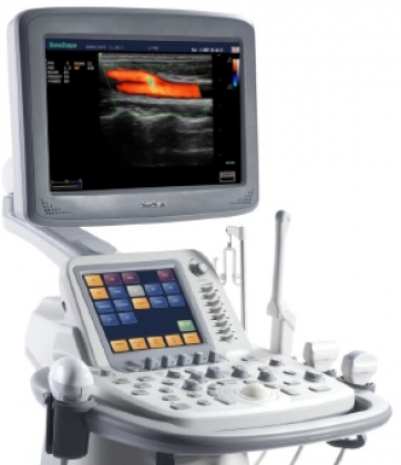
• 17″ high definition LCD monitor with large imaging area
• smart touch screen
• Four transducer sockets for a wide range of transducers: Convex, Phased, Linear, Transvaginal, Transrectal, Bi-plane, TEE, Endoscopic, Intraoperative, 4D
• High density transducers with frequency range from 1.9 to 15 MHz
• 200°transvaginal imaging with Temperature-detection technology
• Integrated with state-of-the-art technologies, like μ -scan, multiple-beam processing,IMT, B-Steer, automatic flow volume analysis
• Application: General, Radiology, Cardiology, OB/GYN, Urology, Vascular
• Full patient database solutions: DICOM3.0, AVI/JPG, USB2.0, HDD, DVD, PDF report
Tidak ada produk berkaitan.
Tidak ada produk popular.
Format Pemesanan Via SMS
Kirim format SMS di bawah ke 08987654321
#Nama #Alamat Lengkap #Kode Produk #Jumlah Produk #Ukuran Produk
Selanjutnya kami akan membalas SMS anda dengan rincian total belanja anda yang harus ditransfer ke rekening kami
Harap segera konfirmasi jika pembayaran sudah ditransfer ke rekening di bawah ini.
