
IMPORTIR & DISTRIBUTOR ALAT LABORATORIUM & ALAT KESEHATAN
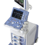
The ProSound Alpha 6 brings outstanding image quality, award winning design, unprecedented feature-rich performance, and ease of use to the office market.
Building on the ProSound technology strengths, this compact system offers high-end features and technologies in an easy-to-use, affordable and environmentally friendly system. The ProSound Alpha 6’s high power processor allows a number of imaging modes previously seen only in high-end systems. The system supports a full range of probes and software, designed to support better diagnostics and patient care. Additionally, the ProSound Alpha 6 is fully upgradeable and easy-to-use.
The lightweight ProSound Alpha 6 has the smallest footprint in its class offering outstanding mobility. With reduced power consumption, the ProSound Alpha 6 is also designed to be economically and environmentally friendly.
Clear images and advanced functions are what you have come to expect from Hitachi – Aloka. Our 60 years of experience and innovation continues with the ProSound Alpha 6 platform.


Real-time 3D allows for the simple and rapid acquisition, optimization, manipulation and analysis of structures in 3D and Real-time 3D/4D.

Creates multiple Multi-planar reconstruction (MPR) images from a 3D volume data and displays the images simultaneously. The MPR images are displayed parallel to each other and assist in the understanding of the internal structure of the 3D image by acquiring the information of multiple planes in a structure.

3D acquisition that provides images of multiple imaging planes including a third plane not accessible with conventional 2D imaging.

Real-time 3D allows the acquisition, optimization, manipulation and analysis of structures in 3D and Real-time 3D/4D

Displays blood flow with directional information at higher frame rates and spatial resolution compared to conventional methods. Detail and accuracy of blood flow information is greatly increased with reduced blooming of color.

Displays blood flow with directional information at higher frame rates and spatial resolution compared to conventional methods. Detail and accuracy of blood flow information is greatly increased with reduced blooming of color.

Real-time 3D allows the acquisition, optimization, manipulation and analysis of structures in 3D and Real-time 3D/4D

3D acquisition that provides images of multiple imaging planes including a third plane not accessible with conventional 2D imaging.

Creates multiple Multi-planar reconstruction (MPR) images from a 3D volume data and displays the images simultaneously. The MPR images are displayed parallel to each other and assist in the understanding of the internal structure of the 3D image by acquiring the information of multiple planes in a structure.

Tidak ada produk berkaitan.
Tidak ada produk popular.
Format Pemesanan Via SMS
Kirim format SMS di bawah ke 08987654321
#Nama #Alamat Lengkap #Kode Produk #Jumlah Produk #Ukuran Produk
Selanjutnya kami akan membalas SMS anda dengan rincian total belanja anda yang harus ditransfer ke rekening kami
Harap segera konfirmasi jika pembayaran sudah ditransfer ke rekening di bawah ini.
