IMPORTIR & DISTRIBUTOR ALAT LABORATORIUM & ALAT KESEHATAN
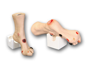
The newest and most comprehensive model of its kind, molded from an 80-year old patient for a true-to-life experience when assessing the various wounds. Twenty different conditions are presented on ‘Wilma’ Wound Foot™ so you can see and understand how they are different.
The newest and most comprehensive model of its kind, molded from an 80-year old patient for a true-to-life experience when assessing the various wounds. Twenty different conditions are presented on ‘Wilma’ Wound Foot™ so you can see and understand how they are different. Great care has been taken to color each wound just as you would see it on a patient. Once the different etiologies are understood, you can discuss and devise treatment plans that will deliver optimized patient care.
Wound assessment has become critical as inaccurate wound assessment can misdirect the plan of care, affect reimbursement, cause inaccurate reporting of patient outcomes and the appearance of potential adverse events. This model is an effective tool for educating all healthcare professionals and patients in the identification and staging of wounds and their probable etiologies. ‘Wilma’ Wound Foot™ is also an excellent visual aid for educating those who cannot read well enough to understand basic health care information, allowing them to see what can occur without proper care. Routine cleansing and dressing changes can be taught and practiced on all the wounds by healthcare providers, patients, families and caregivers.
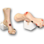
The model is made of flexible material permitting the toes to be moved for closer examination or the application of dressings. This unique material permits the application and removal of dressings without leaving an “adhesive residue”, when used according to supplied instructions. Each model is supplied with a positioning base for the foot which will give “hands free” access to all the sites when applying dressings or for teaching. An optional carrying case is also available.
A great tool for training, competency testing, skills assessment and dressing techniques!
The following wounds*, pressure ulcers* and foot abnormalities are presented on ‘Wilma’ Wound Foot™ :
Size: 12 1/2” X 8” X 3 5/8” Sh. wt. 4.0 lbs.
Tidak ada produk berkaitan.
Tidak ada produk popular.
Format Pemesanan Via SMS
Kirim format SMS di bawah ke 08987654321
#Nama #Alamat Lengkap #Kode Produk #Jumlah Produk #Ukuran Produk
Selanjutnya kami akan membalas SMS anda dengan rincian total belanja anda yang harus ditransfer ke rekening kami
Harap segera konfirmasi jika pembayaran sudah ditransfer ke rekening di bawah ini.
